Ct Scan Brain Anatomy : Anatomy Of Head Ct Scan Normal The Brain On Ct And Mri ... / Frontal sections can be obtained with some devices either directly or by reconstruction ;
Ct Scan Brain Anatomy : Anatomy Of Head Ct Scan Normal The Brain On Ct And Mri ... / Frontal sections can be obtained with some devices either directly or by reconstruction ;. Multidetector ct scans were acquired following the protocol outlined by dupas et al.5 after a lateral topography. Brain, bones of cranium, sinuses of the face. Anatomy ct axial brain form no 4. How to read ct scan. Learn the deep structures of the brain demonstrated by ct scan of the head.
They reveal the brain's anatomy, including the integrity of brain structures and their interconnections. 20 public playlists includes this case. Documents similar to ct brain anatomy. Brain bones of cranium sinuses of the face. Proceedings of the 46th national congress of anesthesiology and reanimation, paris, france.
Interactive CT and MRI Anatomy App Ranking and Store Data ... from static-s.aa-cdn.net Normal brain anatomy as demonstrated by computerized tomography (ct scanning). This means that the right side of the brain is on the left side of the viewer. In a standard scan, the patient is lying with his or her back to the table. Brain ct scans may be done with or without contrast. contrast refers to a substance taken by mouth or injected into an intravenous (iv) line that. Smart practice ensures that you spend more time practicing the questions where you made mistakes. Learn more about ct scans and how to be prepared. Computed tomography (ct) scans are an extremely common imaging modality. Brain bones of cranium sinuses of the face.
Interactive anatomy atlas these pictures of this page are about:ct scan brain anatomy.
The ct head scan is one of the most common imaging studies that you can be faced with and the most frequently requested by a&e. Solve your problem quick & easy with online consultation. Focal abnormalities are not observed in the brain parenchyma. Learn the deep structures of the brain demonstrated by ct scan of the head. A computed axial tomography scan (also called a cat or ct scan) is a frequently tly used technique in brain imaging. Learn about brain anatomy as seen on ct images of the brain. Anatomy ct axial brain form no 4. The most common cause of subarachnoid haemorrhage is trauma, however, they can. Acute subdural hematoma can evolve over a period of time and thus classified as acute, subacute and chronic hematoma. Ct scan hitachi scenaria 128 slice: Frontal sections can be obtained with some devices either directly or by reconstruction ; In a standard scan, the patient is lying with his or her back to the table. Stroke is of two types.
Ct images of the brain are conventionally viewed from below, as if looking up into the top of the head. In a standard scan, the patient is lying with his or her back to the table. Anatomy of the head on a cranial ct scan : Ct brain basics and anatomy by dr.sathish regained! In this article, we will outline the basic principles behind ct scans.
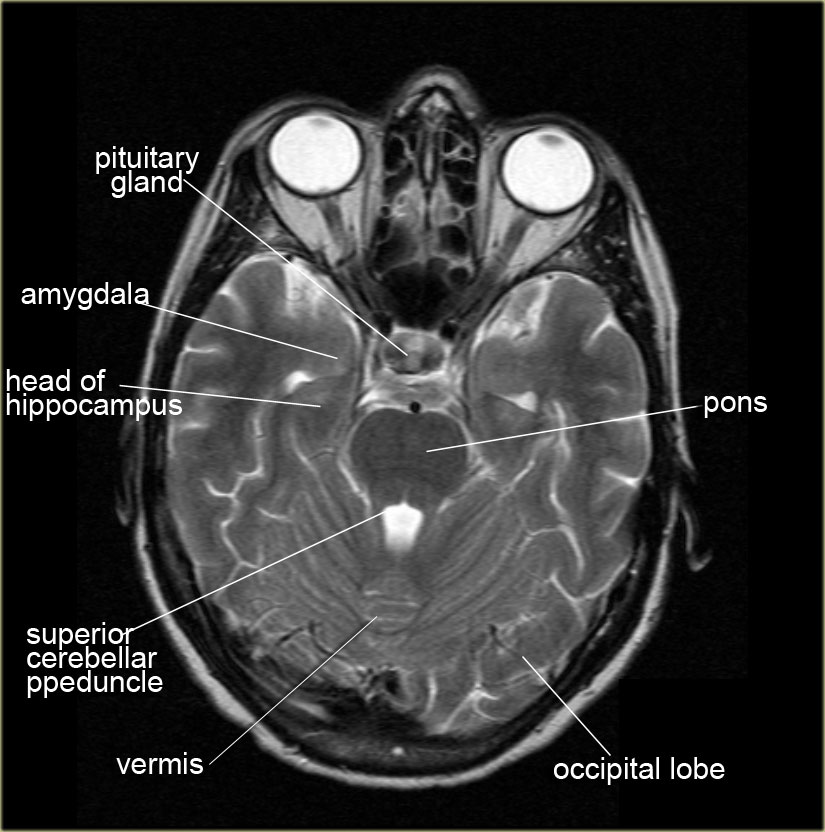
Ct Scan Anatomy Of Brain - Anatomy Drawing Diagram from radiologyassistant.nl Focal abnormalities are not observed in the brain parenchyma. Depending on the scanner, transversal images may be reconstructed in the. Dural venous sinuses, veins, arteries. Proceedings of the 46th national congress of anesthesiology and reanimation, paris, france. Mri and ct are used to confirm the anatomy of stroke and the interpretation of those. The video shows the basic ct anatomy of the brain.for each slice we have highlighted. It contains information about the normal anatomy and the different types of brain hemorrhage. Interactive anatomy atlas these pictures of this page are about:ct scan brain anatomy.
This interactive module will teach ct head interpretation.
Anatomy ct axial brain form no 4. A class discussing the basics of the ct brain examination. Sagittal slices are obtained only by reconstruction. The cerebellum, latin for little brain. Normal brain anatomy as demonstrated by computerized tomography (ct scanning). Smart practice ensures that you spend more time practicing the questions where you made mistakes. Documents similar to ct brain anatomy. The most common cause of subarachnoid haemorrhage is trauma, however, they can. Learn about brain anatomy as seen on ct images of the brain. They reveal the brain's anatomy, including the integrity of brain structures and their interconnections. This means that the right side of the brain is on the left side of the viewer. Learn the deep structures of the brain demonstrated by ct scan of the head. Focal abnormalities are not observed in the brain parenchyma.
Focal abnormalities are not observed in the brain parenchyma. Brain ct scans may be done with or without contrast. contrast refers to a substance taken by mouth or injected into an intravenous (iv) line that. In this article, we will outline the basic principles behind ct scans. Introduction to neuroimaging by keith johnson and alex becker. This interactive module will teach ct head interpretation.
The Radiology Assistant : Brain Anatomy from rad.desk.nl Brain bones of cranium sinuses of the face. Is ct scan brain anatomy your major concern? A class discussing the basics of the ct brain examination. Dural venous sinuses, veins, arteries. It contains information about the normal anatomy and the different types of brain hemorrhage. This means that the right side of the brain is on the left side of the viewer. In a standard scan, the patient is lying with his or her back to the table. Focal abnormalities are not observed in the brain parenchyma.
Ct scans of the brain can provide detailed information about brain tissue and brain structures.
Brain ct scans may be done with or without contrast. contrast refers to a substance taken by mouth or injected into an intravenous (iv) line that. Brain bones of cranium sinuses of the face. Most present devices allow horizontal sections ; The most common cause of subarachnoid haemorrhage is trauma, however, they can. Computed tomography (ct) scans are an extremely common imaging modality. In a standard scan, the patient is lying with his or her back to the table. Documents similar to ct brain anatomy. This article will this space normally contains csf and the vasculature of the brain. The ct head scan is one of the most common imaging studies that you can be faced with and the most frequently requested by a&e. Ct scan hitachi scenaria 128 slice: Learn the deep structures of the brain demonstrated by ct scan of the head. In this article, we will outline the basic principles behind ct scans. Solve your problem quick & easy with online consultation.
You have just read the article entitled Ct Scan Brain Anatomy : Anatomy Of Head Ct Scan Normal The Brain On Ct And Mri ... / Frontal sections can be obtained with some devices either directly or by reconstruction ;. You can also bookmark this page with the URL : https://saripepaya11.blogspot.com/2021/05/ct-scan-brain-anatomy-anatomy-of-head.html
Share Awesome
Belum ada Komentar untuk "Ct Scan Brain Anatomy : Anatomy Of Head Ct Scan Normal The Brain On Ct And Mri ... / Frontal sections can be obtained with some devices either directly or by reconstruction ;"


Belum ada Komentar untuk "Ct Scan Brain Anatomy : Anatomy Of Head Ct Scan Normal The Brain On Ct And Mri ... / Frontal sections can be obtained with some devices either directly or by reconstruction ;"
Posting Komentar